
Special Equipment
Equipment Used in the CRC Lab
RTCA SP Station
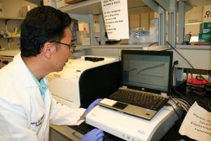
The RTCA SP Station is located inside a tissue culture incubator and is capable of switching any one of the wells on the E-Plate 96 to the RTCA Analyzer for impedance measurement. Under the software of the RTCA Control Unit, the RTCA SP Analyzer can automatically select wells for measurement and continuously transfer measured impedance data to the computer. Cell Index values, derived from the measured impedances, are continuously displayed on the Software user interface.
IncuCyte Zoom® Live Cell Imaging
IncuCyte ZOOM® enables observation and quantification of cell behavior over time by automatically gathering and analyzing images around the clock. All within the controlled environment of a standard cell incubator.IncuCyte ZOOM® consists of a microscope gantry that resides in the cell incubator and a networked external controller hard drive that gathers and processes image data. Different microscope objectives (4x, 10x, 20x) can be housed within each system and readily interchanged by the user. Each IncuCyte ZOOM® houses multiple T-flasks or microtiter plates (up to 6) and can acquire >2000 images per hour.
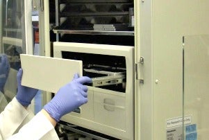
Variable Oxygen Control Incubators
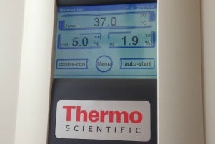
Inside the body, oxygen concentrations range from 1-14%, rather than the 20-21% found in the atmosphere. Cells cultured in low oxygen (hypoxia) grow faster, live longer, and show lower stress. Our variable oxygen control incubator will generate hypoxic conditions (around 2% oxygen, with no changes in humidity and CO2 level) to help your cells grow faster and healthier.
Microscope: EVOS® XL Cell Imaging System
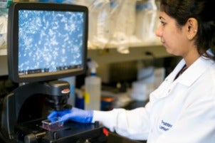
Cell Imaging System is an instrument for transmitted-light microscopy, colorimetric applications like H&E staining and immunohistochemistry, as well as time-lapse microscopy.